Iranian Firm Manufactures Fluorescent Molecular Imaging Device
This advanced system, designed for non-invasive research on animal models, provides a new perspective in oncology, immunology, and cardiovascular research with capabilities like high sensitivity, multi-wavelength imaging, and live video recording.
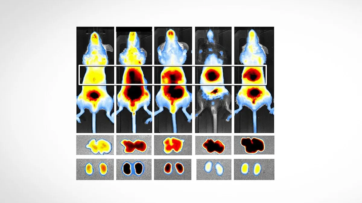
FluoVision, which is offered at a much lower price than foreign models, allows researchers to track the accumulation of biological molecules and observe lymph nodes without the need for complex cooling and with user-friendly software, and reduces dependence on imported technologies.
The FluoVision molecular imaging device provides a powerful tool for two-dimensional fluorescent imaging in vivo and on laboratory animal models.
The FluoVision device is designed to track and study biological processes at the molecular level. Its most important technical features include very high sensitivity in detecting low light intensities, the ability to image at different wavelengths, and advanced filtering capabilities for precise separation of emitted signals.
This system uses flexible light sources that can simultaneously install 4 light sources with different wavelengths (usually 460, 485, 530, and 740 nm), which can also be customized based on the researcher's needs. On the other hand, 5 optical filters (usually 500, 550, 600, 650, and 800 nm) can also be installed, which greatly increases the accuracy of detection. One of the engineered advantages of this device is the lack of the need for external cooling, which has made its use and transportation very simple.
In a relevant development in December, an Iranian knowledge-based company stationed at Pardis Science and Technology Park had also managed to produce an atomic force microscope which is used to image and characterize samples at the nanoscale.
“One of our knowledge-based products is the atomic force microscope, which is used to image and characterize samples at the nanoscale. This device uses a sharp-pointed probe to scan the sample surface. During the scanning operation, laser light is emitted to the back of the cantilever and its reflection on a photodiode will result in the formation of an image of the sample surface,” said Seyed Abbas Shahmoradi, the managing director of the knowledge-based company.
He mentioned the Bio-AFM as another achievement of his company, and said, "This microscope is one of the most important tools for studying samples in biology, because Bio-AFM provides a suitable platform for integrating atomic force microscopy and optical microscopy in biological research projects."
Noting that the Bio-AFM microscope can capture images in different environments with diverse working modes, Shahmoradi said, “This capability allows scientists to study the structure and properties of living cells and other biological samples like DNA and RNA, proteins, viruses, bacteria, and tissues."
“Also, the Transmission Electron Microscopy (TEM) produced by our company, with an accuracy of 0.6 nanometers, is capable of providing images with extremely high resolution. For comparison, the size of the coronavirus is 120 nanometers, while the accuracy of our device is one two-hundredth of this value,” he added.
4155/v


