Novel Eye Scanner Technology Targets Neurodegenerative Diseases

In a large-scale EU project, in which MedUni Vienna has collaborated with multiple international partners, the capabilities of optical coherence tomography (OCT) and Raman spectroscopy have been utilised to create a novel eye scanner. This new technology can combine molecular information to the visualisation of the internal structure of the eye. This has the potential to enable the early detection of neurodegenerative diseases, as well as other eye diseases and diabetes, the Innovation News Network reported.
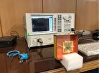
A multimodal eye scanner was developed as part of the MOON project. This scanner not only provides a high-resolution image of the structure of an eye using OCT, but also delivers sensitive molecular characterisation of the tissue. To achieve this, the capabilities of OCT have been combined with those of Raman spectroscopy.
The latter technology uses light to detect the finest molecular vibrations, allowing the molecules in the tissue to generate a characteristic spectrum, from which it is possible to determine the composition of the tissue. By combining the expertise of the MOON project partners, it was possible to develop the first multimodal device that provides both OCT and Raman spectroscopic data from the living human eye.
Biochemical changes due to disease-related processes occur long before there is actual tissue damage, which can result in an irreversible loss of vision, especially in the retina. The earlier such changes are detected, the better for the patient, as most treatments cannot reverse existing damage but rather aim to stop its progression.
Early diagnosis of disease therefore encompasses both spectroscopic and functional identification of tissue status, in addition to structural imaging. The image quality and image size of functional and structural information achieved with the novel eye scanner technology are unrivalled anywhere in the world. The use of Artificial Intelligence allows additional contrast enhancement of OCT angiographs for functional imaging.
A study, currently being conducted at the Department of Ophthalmology and Optometry of the Medical University of Vienna, highlights the relevance of advanced OCT technology for improved ocular diagnostics and treatment planning in diabetic patients. With these results, the Medical University of Vienna is establishing new standards in retinal diagnostics based on OCT alone.
Ongoing clinical trials with researchers in the field of ophthalmology, neurology, nuclear medicine, and clinical pharmacology at the Medical University of Vienna are not only investigating eye diseases but also neurodegenerative diseases. Based on the group’s hypothesis, they predict that neurodegenerative diseases of the brain can also lead to changes in the sensitive retinal nerve tissue, meaning the eye can serve as a window to the brain.
Alzheimer’s disease is being studied as an important example of this type of neural disease. In the ongoing clinical investigations, the very first relevant Raman spectra from the human eye has now been recorded, with initial indications of its diagnostic potential.
Further studies are already scheduled, with clinical partners from ophthalmology and clinical pharmacology at the Medical University of Vienna. These will explore the validity of Raman spectroscopy in the diagnosis of diabetes and neurodegenerative diseases other than Alzheimer’s disease such as Multiple Sclerosis or Parkinson’s disease.
4155/v


