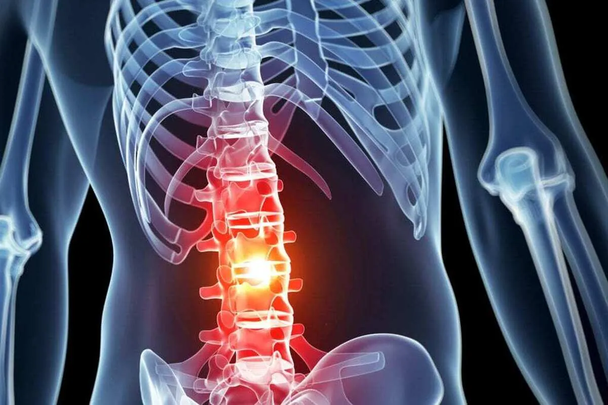
Iranian Scientists Gain New Achievement in Tissue Engineering, Treatment of Spinal Cord Injuries
The research project titled ‘investigating the anti-inflammatory effects of scaffolds containing quercetin nanoparticles in a unilateral spinal cord injury model in male rats’ was carried out by Gholamreza Hassanzadeh, the professor of the department of Anatomical Sciences and the director of the Department of Neuroscience and Addiction at Tehran University of Medical Sciences.
The research was conducted with the aim of developing new strategies for the treatment of spinal cord injuries and was able to reveal new horizons in the treatment of spinal cord injuries by investigating the role of sustained secretion of quercetin in reducing inflammation.
“The results of the study showed that continuous secretion of quercetin can help improve the recovery of patients with spinal cord injuries and be of interest to researchers and physicians as a new approach,” Hassanzadeh said.
“This achievement can encourage researchers in preclinical and clinical fields to utilize scaffolds containing quercetin nanoparticles. Also, if the effectiveness of this method is confirmed in more advanced studies, it will pave the way for its introduction as a new treatment protocol for spinal cord injury patients,” he added.
In a relevant breakthrough in 2024, a group of Iranian researchers from Shiraz University of Medical Sciences could find a way to regenerate nerve tissues and spinal cord injury by extracting mouse neural stem cells and using ibrutinib drug.
“In this project, we studied the combined effect of stem cells of the nervous system and drug ibrutinib (targeted drug for cancer treatment) to regenerate nerve tissue and recovery of motor function in a mouse with spinal cord injury,” Somayyeh Torabi, a researcher of the project, told ANA.
“In this research, we used the 14-day-old mouse embryo’s stem cells; a region of the brain of this mouse is rich in stem cells, and these cells are easily harvested and extracted,” she added.
“After we extracted stem cells from these embryos, we cultured them. To do this, we did initial cultivation. Finally, after 6-7 days, the cells started multiplying, and after a month, we transplanted these cells into the spinal cord lesion models,” Torabi said.
Noting that these cells were used for adult mice in which they created a spinal cord lesion, she explained, “We injected these cells in the area where the lesion was created. Then we monitored and observed the animals for a month. We tested their sensory, motor and balance performance. At the end of the month, we checked their tissue repair.”
“In this project, we measured the size of the lesions and the behavior of the transplanted cells in different groups, because along with the cell transplant, we also used an anti-inflammatory drug of the immune system. We also investigated this issue to observe the behavior of the transplanted cells in the target tissue - which had received drugs or not -. Finally, we obtained an acceptable result of the survival and fate of the cells,” Torabi said.
4155/v


