Scientists Crack Mystery of Abnormal Protein Accumulation

Nucleotides are the basic building blocks of DNA and RNA. DNA and RNA each contains four bases, namely adenine (A), cytosine (C), guanine (G), and thymine (T in DNA) or Uridine (U in RNA). Sometimes genes may contain segments of several nucleotides that repeated many times, called microsatellite repeat expansion. Once the expansion is too much, it will cause some neurological diseases, the journal Nature reported.
Previous research showed that longer CAG (Cytosine, Adenine, Guanine) repeat lengths in RNA are associated with earlier onset and more severe neurodegenerative diseases.
In the study, the research team led by Wang first discovered that the A in the CAG repeat RNA are methylated into N1-methyladenosine (m1A), and the degree of methylation in the CAG repeat increased significantly with the increase of the number of repeats.
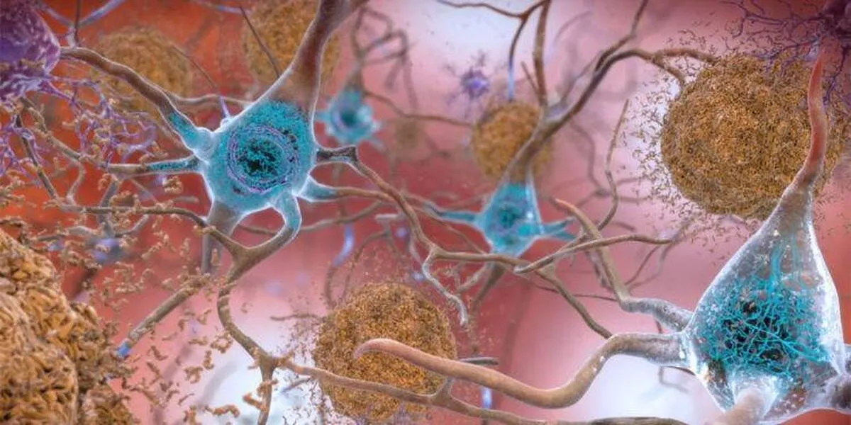
Further, m1A directly bind to TDP-43 (a DNA-RNA binding protein normally found in the nucleus with molecular weight 43KD), causing the protein to mis-localize to the cytoplasm instead and form gel-like aggregates, which is consistent with protein accumulation abnormalities observed in neurodegenerative diseases.
m1A methylation is catalyzed by m1A methyltransferase, a heterodimer enzyme complex, in which the highly conserved TRMT61A, from invertebrates to humans, is the catalytic subunit of the methyltransferase complex.
The authors revealed that TRMT61A is required for the A in the CAG repeat RNA to be methylated to m1A, and the enzyme ALKBH3 is responsible for the demethylation of m1A back to A.
To study the impact of m1A in CAG repeat expansion RNA on neurodegenerative diseases, the researchers constructed a Caenorhabditis elegans model (Q67) of neurons with 67 CAG repeats, and a Drosophila model (Q78) with 78 CAG repeats.
Both the neural network of Q67 and Q78 display significant degeneration. However, in transgenic C. elegans expressing human ALKBH3, the signs of neural degeneration are reduced, while C. elegans expressing a catalytically inactive mutant human ALKBH3 did not produce these improvements. This suggests that m1A promotes the neurotoxicity of CAG repeat RNA, reducing the m1A fraction mitigates the toxic effect.
In order to figure out how m1A promotes neurotoxicity, the researchers screened several m1A-binding proteins, including TDP-43, YTHDF1-3 and DDX56. Among them, TDP-43 bound to m1A the most, had the strongest binding ability, and preferentially m1A binding in CAG repeat RNA in cells.
Structurally, TDP-43 contains two RNA recognition motifs (RRMs), in which highly conserved aromatic amino acid residues are required for their interactions. m1A was shown to induce cytoplasmic mis-localization of TDP-43, and when co-cultured, TDP-43 and m1A combine to facilitate a liquid-to-gel-like transition of TDP-43. m1A in CAG repeat RNA also promotes the sequestration of TDP-43 into stress granules in cells.
These results revealed a novel pathological function of m1A - in binding with TDP-43 and eliciting its aberrant biochemical and biophysical properties, which explains the role of this protein in many neurological diseases.
It is noted that neurotoxicity observed in CAG repeat expansion disorders may also arise from other mechanisms, e.g. polyglutamine-containing proteins translated from the CAG repeat-bearing RNAs, and the following perturbation of the ubiquitin-proteasome system. Thus, multiple mechanisms probably contribute simultaneously to neurodegenerative diseases caused by nucleotide repeat expansions.
The new pathological function of m1A in CAG repeat RNA suggests for new therapies to treat neurodegenerative diseases. Specifically, the identification of the two enzymes, methyltransferase (TRMT61A) and demethylase (ALKBH3) for m1A, provides a strong molecular foundation for such future efforts. Treatments that target methylation/demethylation process of m1A may have the therapeutic potential to reduce neurodegeneration and increase life span for patients.
4155/v


