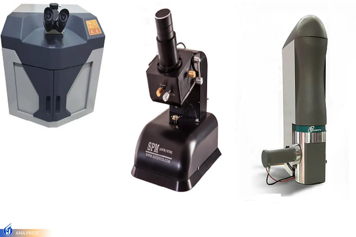
Nano Laboratories in Iran Equipped with Indigenized Modern Microscopes

One of the technological companies in Iran has produced and commercialized scanning electron microscope (SEM) microscopes with the capability to study morphology, chemical composition of the surface and determine the thickness of thin layers coated on a substrate.
This electron microscope enjoys a resolution of 20 nm and has a maximum magnification of 100,000 times, and its tungsten thermal electron gun makes imaging different materials possible.
Another Iranian firm active in the field of nanotechnology, which has more than a decade of experience in the production and supply of Scanning Tunneling Microscope (STM) and Atomic Force Microscope (AFM) microscopes, has made microscopes for research purposes as well as special microscopes for the education sector.
The Scanning Probe Microscopy (SPM) equipment of this company provides the possibility of 3D imaging of the topography of samples to study indicators like roughness, grain size, step height and unevenness, and also gives interesting details about other sample specifications like magnetic field, capacitance, friction and phase.
The researchers of another company in Iran have produced and commercialized a Raman microscope for identifying molecules and studying chemical bonds, which has a spectral range between 200 nm and 900 nm and a spectral resolution of 1.3 nm. This Raman spectrometer can be used to study a wide range of nanoparticles to final products like various polymer nanocomposites.
Another Iranian company's engineers have developed special transmission electron microscope (TEM) cameras that several research centers and universities are using to improve the performance of their TEM microscopes. These cameras have improved the ability of the TEM microscope to provide better images and also increased their imaging speed.
In a relevant development earlier this year, researchers at a knowledge-based company in Iran succeeded in producing microscopes whose output is 3D images used for verification or different experiments.
“The researchers of our company succeeded in designing a microscope that provides the user with a 3D image of the sample in addition to enjoying the efficiency of other microscopes,” Massoumeh Nasseri, the sales manager of the company, told ANA.
Noting that the microscope takes images of the sample at different zooms, she said, “These 3D images have different uses for verification and various experiments.”
“Banks, judiciary, research centers, universities, document registration offices and institutions that are dealing with testing can use this microscope,” Nasseri emphasized.
She added that no sample of the microscope has yet been made in Iran, stating that there are no foreign models either.
Nasseri had also last year announced manufacturing of an AFM for imaging living biological samples.
4155/v


