New suturing method aims to reduce cesarean complications
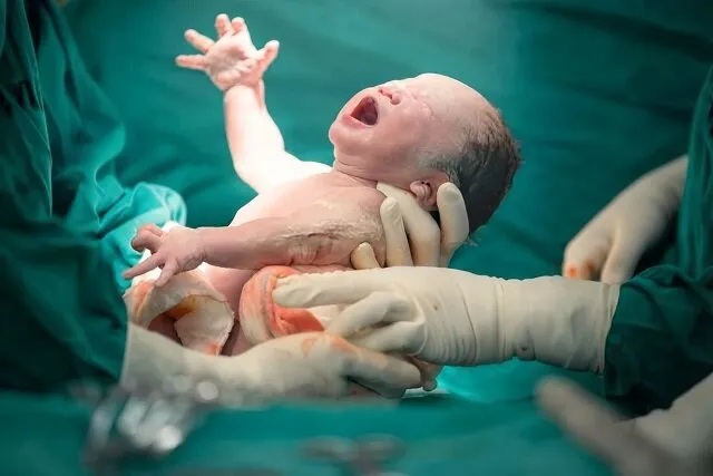
Cesarean section is one of the most common surgical procedures worldwide. It is performed when medically indicated to protect the health of the mother or baby, or at the mother’s request. Despite its widespread use, the procedure can be associated with significant complications, including severe bleeding during or after delivery, localized blood clot formation, adhesions between internal abdominal organs and painful infections.
Other potential complications include abnormal scar formation and weakening of the uterine wall at the incision site, a condition known as a cesarean scar defect or cesarean scar niche. These abnormalities may increase the risk of serious complications, including uterine rupture in subsequent pregnancies, particularly when attempting vaginal birth after cesarean delivery.
The technique used to suture and close the uterine wall after the delivery of the baby and placenta plays a crucial role in determining recovery time, postoperative pain, the risk of short- and long-term complications, and maternal safety.
Conventional approaches, including single-layer and double-layer uterine closure, as well as more complex techniques, have various limitations. Some place excessive tension on tissues, potentially restricting blood flow, causing ischemia and increasing tissue damage and scar formation. Others prolong surgery or fail to adequately cover tissue layers, increasing the risk of adhesions between abdominal organs.
Developing a technique that preserves the uterus’s natural anatomy while promoting more precise healing and reducing complications is therefore an important clinical objective.
Against this backdrop, Nayera Seyedi-Moghadam, together with colleagues at the Research Center for Obstetrics, Gynecology and Infertility and Sarem Women’s Hospital, conducted an applied study introducing a modified, specialized technique for suturing uterine tissue during cesarean section.
In the proposed method, uterine repair is performed in several carefully controlled stages. For the first layer, suturing begins at one corner of the uterus using standard size 0 Vicryl surgical thread. The uterine muscle, or myometrium, is then closed with a continuous, non-locking suture extending to the other end of the incision.
For the second layer, suturing begins at the opposite corner of the uterus. To preserve its natural structure and anatomy, part of the muscular tissue, together with the outer covering known as the uterovesical fascia, is sutured continuously without locking, using the same type of thread.
Finally, to reinforce tissue repair and prevent the uterus from adhering to surrounding abdominal tissues, the peritoneal layer is sutured separately using finer size 2-0 Vicryl thread.
The study’s findings suggest that the technique may offer several improvements in postoperative recovery. By suturing each layer separately, the method allows the different tissue layers to heal independently and naturally, potentially reducing postoperative inflammation and pain.
The researchers also reported improved bleeding control, a lower risk of uterine rupture in future deliveries and fewer structural defects at the surgical incision site.
The study concludes that the modified suturing technique may offer several advantages over conventional methods and other approaches, including better control of bleeding, shorter hospital stays, faster recovery and an earlier return to daily activities. By reducing tissue damage and abdominal adhesions, it may also help preserve women’s future reproductive health. However, larger clinical studies are still needed to confirm these findings.
Further details published in the Sarem Medical Research Quarterly, affiliated with Sarem Women’s Hospital, explain the limitations of existing approaches. In conventional single-layer closure, incomplete bleeding control may lead to increased blood clot formation beneath the tissue. Excessive tension may also increase postoperative pain and the risk of uterine rupture in future pregnancies.
By contrast, the new technique uses minimal tension and carefully aligns the tissue layers, helping to limit tissue damage and severe pain.
Conventional double-layer techniques often rely on locking sutures that place considerable tension on the tissue and may compromise its blood supply. Failure to suture the peritoneal layer separately can also increase the risk of adhesions between internal organs.
The modified approach addresses these limitations through non-locking sutures and separate closure of the peritoneum. According to the researchers, this combination may reduce adhesions and improve uterine wall healing while avoiding some of the time-consuming and costly aspects of other techniques, striking a balance between surgical efficiency and effectiveness.