AI algorithms detect cancer cells circulating in blood
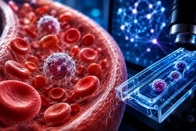
Circulating tumor cells (CTCs) are cancer cells that break away from a primary tumor and enter the bloodstream. Their presence can indicate that cancer may be spreading through the body. Detecting these rare cells could help with early diagnosis, monitoring disease progression and assessing treatment effectiveness.
However, identifying CTCs is challenging because they are extremely rare compared with other blood components. A blood sample may contain millions of red and white blood cells, while CTCs can be present in very small numbers. This makes their detection and counting difficult and time-consuming.
Various techniques have been developed to isolate CTCs, including the use of specific antibodies, separation based on cell size or density, analysis of cell adhesion, and physical properties such as acoustic and hydrodynamic characteristics. Each approach has advantages and limitations, highlighting the need for more accurate and efficient methods.
Against this backdrop, Hajar Moghaddas, an assistant professor in the Department of Mechanical Engineering at Yasouj University, and a university colleague conducted a review of the use of artificial intelligence to detect CTCs in blood samples.
The researchers systematically examined previous studies on the application of machine learning and deep learning for the automated identification of cancer cells in real blood samples. The review assessed the advantages and limitations of different approaches, including their high speed and accuracy as well as their dependence on the quality and volume of training data.
The findings showed that AI model performance depends heavily on image quality. Clearer, more detailed images allow algorithms to distinguish cancer cells from other blood cells more accurately. However, training these models can be time-consuming and requires large datasets.
To address this challenge, researchers have proposed techniques such as image preprocessing, noise reduction and synthetic data generation. Increasing the number of training images through techniques such as rotation, magnification and rescaling can also improve model performance.
The review found that the AI models examined generally achieved greater speed and accuracy than conventional methods for counting and classifying cancer cells. Nevertheless, further development is needed before these approaches can be routinely used in clinical settings, particularly models capable of performing reliably with limited data and across different laboratory conditions.
The researchers also noted that removing irrelevant parts of images can speed up processing. However, excessive manipulation of blood samples, including the complete removal of certain blood components, could result in the loss of cancer cells and lead to inaccurate results.
One proposed approach is to use diluting agents to reduce particle density in images, making cell boundaries easier to identify. This strategy, however, may increase the number of images required for analysis.
The study, whose findings were published in the Journal of Mechanical Engineering of the Iranian Association of Mechanical Engineers, concludes that AI has considerable potential to address challenges in detecting and counting circulating cancer cells and could contribute to faster and more accurate diagnostic methods in the future.